
Knobology and Image Optimisation
- Description
- FAQ
- Reviews

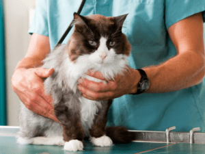
Ultrasound is one of the most powerful diagnostic tools in small animal practice, but only if you know how to get the best from your machine. In this free introductory course, Dr Caroline Taylor BSc (Hons) BVetMed PGCert(SAM) FHEA MRCVS guides you through the essentials of ultrasound image optimisation—also known as Knobology.
Across twelve short lessons, you’ll explore the science behind ultrasound image formation, how to prepare your patient and set up the scanning environment, and how to master the “Five Magic Buttons” that make the biggest difference to image quality. You’ll also learn to recognise and correct common artefacts, giving you the confidence to produce consistently clear and diagnostic images.
Designed with both veterinary surgeons and registered veterinary nurses in mind, this module is a practical, interactive starting point for anyone wanting to strengthen their ultrasound skills before moving on to more advanced abdominal or thoracic scanning.
By the end of the course you will:
- Understand the physics behind ultrasound image creation.
- Apply patient preparation and probe handling techniques to optimise image quality.
- Confidently adjust key machine controls (Depth, Gain, Focus, Time Gain Compensation, Frequency).
- Recognise common ultrasound artefacts and know how to respond to them.
This course is the foundation for the Skill Vet Scan Point-of-Care Ultrasound pathway and will prepare you for further training in abdominal, thoracic, and echocardiographic scanning.


-
Format: 12 short, interactive lessons with video, diagrams, and scenario-based questions. Note: This course is best viewed on a desktop computer, laptop or tablet.
-
Length: Approx. 2 hours (self-paced)
-
Instructor: Dr Caroline Taylor BVetMed PGCert(SAM) FHEA MRCVS
-
Includes: Downloadable quick-reference guides on machine controls and artefact troubleshooting
-
Certificate: PeerVet digital certificate of completion
-
Note: This course is best viewed on a desktop computer, laptop or tablet.
-
No formal ultrasound training required.
-
Some prior exposure to ultrasound in practice is helpful (e.g. observing scans or assisting colleagues).
-
Access to an ultrasound machine in practice to apply learning.
-
Veterinary Surgeons (VS): any vet who wants to gain confidence with ultrasound, whether new to imaging or looking to refresh core skills.
-
Registered Veterinary Nurses (RVNs): those supporting diagnostic imaging and wanting to build practical knowledge of ultrasound use.
-
Suitable for both UK and international veterinary professionals.

